Related Article
Many adults brush off tiredness, breathlessness, or a fluttering heartbeat as part of a busy routine. However, minor symptoms like these can be a sign of something more serious, such as heart valve disease. Since this cardiac condition often develops quietly, recognising when something feels different is an important first step.
In this article, we walk you through the essentials: what heart valve disease involves, how it manifests in adults, and the steps you can take to safeguard your heart health.
The heart contains four valves: aortic, mitral, tricuspid, and pulmonic. These valves regulate blood flow by opening and closing in a controlled sequence. Heart valve disease develops when one or more of these valves become narrowed, stiff, or unable to seal properly, straining the heart to maintain circulation.
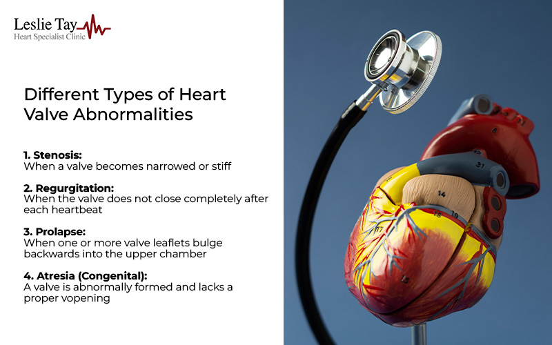
Although heart disease can affect any of the four valves, the way each valve malfunctions tends to fall into predictable patterns. Identifying the correct type helps doctors determine the best management plan.
Stenosis occurs when a valve becomes narrowed or stiff. The valve leaflets may have thickened over time, developed calcium deposits, or fused edges. The opening becomes smaller, and more pressure is needed to push blood through.
Aortic stenosis is one of the more common forms of heart valve disease in older adults. In Singapore, its prevalence is increasing as the population ages, affecting about 3% of individuals over 75 years old and up to 8% of those over 85. It develops slowly over decades, and patients may first notice symptoms like reduced exercise tolerance or chest tightness during exertion.
Regurgitation happens when the valve does not close completely after each heartbeat. This allows blood to leak backwards, reducing the amount of blood pumped forward. The heart compensates by enlarging slightly to accommodate the extra volume, potentially affecting heart muscle strength in the long run.
Mitral regurgitation is the most common heart valve disease in individuals of all ages, affecting over 2% of people globally. It may stem from age-related changes or high blood pressure.
Valve prolapse occurs when one or more valve leaflets bulge backwards into the upper chamber, affecting how tightly the valve closes. If a gap forms, blood can leak backwards through the valve, leading to regurgitation.
Not all cases of prolapse create enough structural distortion to cause leakage, which is why many cases remain harmless. However, when prolapse becomes more pronounced or the supporting structures of the valve weaken, regurgitation may develop, sometimes leading to palpitations, breathlessness, or reduced exercise tolerance.
Valve atresia is a congenital condition in which a valve is abnormally formed and lacks a proper opening. Many people with atresia undergo corrective procedures in childhood, though long-term follow-up is still important, as valve function can change over time and lead to further complications in adulthood.
Heart valve disease is also commonly described as mild, moderate, or severe.
Mild valve narrowing or leakage may not cause symptoms. Many patients continue their usual activities without difficulty. At this stage, monitoring is the key approach, with echocardiograms typically recommended every 12 to 24 months to track any changes in valve function.
Moderate valve disease reflects more noticeable changes in how the valve opens or closes. Some individuals may begin to experience breathlessness, reduced stamina, or palpitations during physical activity. Doctors may recommend more frequent follow-up, often every 6 to 12 months, to monitor for progression.
Severe valve disease indicates a substantial change in valve function, placing greater strain on the heart. Symptoms may occur even with light activity or at rest. At this stage, doctors often discuss options like valve repair or replacement to prevent complications and support long-term heart health.
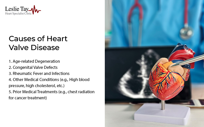
While some causes are well-defined, others relate to a gradual combination of ageing, lifestyle factors, and medical history.
The valves are constantly exposed to pressure changes with every heartbeat. Across decades, the tissue may thicken or accumulate calcium, especially in the aortic valve. Calcium makes leaflets less flexible, limiting valve opening. Stiff tissue may also prevent a tight seal, contributing to regurgitation.
Some valve abnormalities are present at birth but do not cause problems until adulthood. A bicuspid aortic valve, for instance, has two leaflets instead of three. Although it may function normally at first, it can become narrowed earlier in life compared to a typical valve.
Though less common today, rheumatic fever remains a recognised cause of heart valve disease in adults. The inflammation from the illness can leave lasting scarring or deformity, which may lead to stenosis, regurgitation, or a combination of both later in life. Infections such as infective endocarditis can also harm valve tissue, especially when bacteria attach to and weaken the valve surfaces.
Other long-standing medical conditions may also contribute to the development of heart valve disease.
Conditions that affect connective tissues, such as Marfan syndrome, may also stretch or weaken valve structures.
Patients who have received chest radiation for cancer treatment or undergone previous heart surgery may develop heart valve disease years after therapy. Radiation exposure can stiffen or thicken valve tissue gradually.

One challenge with heart valve disease is that symptoms tend to appear slowly. Many adults assume their tiredness or breathlessness relates to stress, fitness level, or ageing.
While some symptoms progress gradually, others may indicate that the heart is under increasing strain:
In more severe cases, sudden shortness of breath, fainting episodes, rapid swelling, or unexplained weight gain from fluid retention should be treated as potential red flags and may require emergency care.
Without monitoring or timely treatment, heart valve disease can gradually place increasing strain on the heart. Over time, this may lead to complications such as:
Early evaluation allows doctors to identify subtle changes before they progress, making it easier to plan treatment proactively. If you notice new or worsening symptoms, consider undergoing a comprehensive heart screening in Singapore. Timely diagnosis of heart valve disease enables doctors to closely monitor the condition, manage symptoms, and plan interventions when necessary.
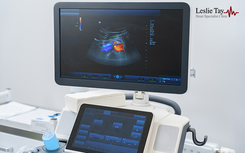
Doctors may recommend a combination of imaging studies, a physical examination, and functional testing. These assessments provide insights into how the valve performs and how the heart compensates for changes in blood flow.
An echocardiogram is one of the primary tools used to diagnose heart valve disease. It is used to obtain high-resolution images of valve structure, measure blood flow direction and speed, and assess heart chamber size and pumping function.
During the examination, a small probe will be placed on your chest to capture images of your heart and valves. Patients often report hearing soft pulsing sounds as the machine measures blood flow. Since the procedure is non-invasive, it is often used for both initial diagnosis and ongoing monitoring.
An ECG test cannot diagnose valve disorders on its own, but it provides important information about the heart’s function. It can reveal irregular rhythms, signs that the heart muscle has thickened from long-term strain, or other electrical abnormalities.
During the test, small adhesive electrodes are placed on your chest, arms, and legs. You will be asked to lie still for a few minutes while the machine records your heartbeat.
A chest X-ray provides a quick overview of the heart and lungs, helping to identify potential changes or abnormalities. For example, an enlarged heart silhouette can suggest long-standing valve leakage or narrowing, while the presence of fluid in the lungs may indicate that the heart is struggling to maintain normal circulation. The test itself is brief and typically involves standing against an imaging panel while a single picture is taken.
A cardiac MRI offers detailed images of the heart’s structure and function, allowing doctors to assess valve function, measure blood flow, and evaluate the condition of the heart muscle. During the scan, you will lie on a table that moves into the MRI machine. You may hear rhythmic tapping sounds as the images are captured, and you may be asked to hold your breath briefly at intervals. This test does not involve radiation and is often used when more specific information is needed.
Exercise or stress tests assess how the heart responds to physical activity, which can reveal symptoms or changes that may not be noticeable at rest. For instance, breathlessness or chest discomfort early in the test may indicate that valve disease is limiting blood flow.
For exercise-based tests, you may be asked to walk on a treadmill or use a stationary bicycle while your heart rhythm, blood pressure, and breathing are monitored. The activity increases gradually based on your comfort level and medical suitability. Individuals who cannot exercise may be given medication to simulate the effects of activity.
When surgery or valve repair is being considered, doctors may request a coronary angiography to evaluate the condition of the heart’s arteries. During this procedure, a thin catheter is inserted through an artery in the wrist or groin, and contrast dye is used to make the arteries visible on imaging. Local anaesthesia is applied at the insertion site, and patients remain awake as the images are obtained.
Identifying the presence of artery disease is crucial when planning treatment, particularly for older adults or individuals with additional cardiovascular risk factors. Blocked or narrowed arteries may influence the type of procedure recommended and whether additional interventions are needed at the same time.

Valve disease management varies depending on severity, symptoms, age, lifestyle, and overall health. In general, treatment does not follow a one-size-fits-all approach and instead evolves as the condition changes.
Medication cannot completely cure heart valve disease, but it may help manage symptoms or stabilise contributing factors. Doctors may prescribe treatments to:
Medication is typically used when the condition is mild or when symptoms are manageable. Additionally, patients who begin medication will still require regular cardiac testing and follow-up because valve structure may continue to change over time.
Valve repair aims to correct the existing valve rather than replace it. Repair techniques vary depending on the valve and problem involved:
Repair is often considered when the valve’s tissue is still healthy enough to function well after adjustment. For suitable patients, some repairs may be performed using minimally invasive procedures, which involve smaller incisions and less downtime.
When the valve is too damaged to repair, replacement becomes the recommended option. Doctors may discuss two main types of replacement valves:
Replacement may be performed through open-heart surgery or, for certain conditions, through minimally invasive techniques such as Transcatheter Aortic Valve Implantation/Replacement (TAVI/TAVR). This approach is often considered for older adults who are at high risk for open-heart surgery, have moderately severe aortic stenosis, or are above the age of 70–80.
Regardless of whether patients undergo repair or replacement, follow-up care remains essential. This may include:
Patients living with heart valve disease often work closely with their cardiologist to create a long-term plan that supports both heart function and daily activities. Many individuals continue to lead active, meaningful lives with appropriate monitoring and treatment.
No. Once a valve is damaged, it cannot heal naturally or return to normal function without medical intervention. Medications may help control symptoms such as shortness of breath, swelling, or irregular heartbeat, but they do not fix the underlying valve problem. Depending on the severity, long-term management may involve regular monitoring, repair, or replacement to prevent complications.
If left untreated, valve disease can worsen over time and place increasing strain on the heart. This can eventually lead to heart failure, stroke, blood clots, or even sudden cardiac death in severe cases. Many people adapt to mild symptoms and delay treatment, but timely diagnosis and early intervention can help improve both quality of life and long-term survival.
For mild cases, doctors often recommend an echocardiogram every 12 months to track changes in valve function. If the condition progresses or symptoms become more noticeable, monitoring may be required every 3–6 months. The exact frequency depends on the type of valve problem, its severity, and the patient’s overall health, as advised by your specialist.
Not all valve disease can be prevented, especially when caused by age-related wear or congenital defects. However, you can lower your risk by:
Many people with valve disease can exercise safely, but the type and intensity should match the severity of the condition. Light to moderate activity is often suitable for mild disease, while more advanced cases may require adjustments to avoid placing strain on the heart. A specialist can recommend an exercise plan tailored to your fitness level, symptoms, and valve function.
Life expectancy varies depending on age, overall health, the type of procedure performed, and how well the heart functions after treatment. For example, individuals aged 60 to 64 who undergo surgical aortic valve replacement have reported an average life expectancy of around 16 years. Ongoing follow-up care remains important to ensure the repaired or replaced valve continues to function as intended.

Heart valve disease can be easy to miss at first. Symptoms often appear gradually, and many adults adjust their routines without realising that their heart may be working harder than it should. But, with today’s diagnostic tools, structured follow-up, and a range of treatment approaches, there are many options to help manage and treat this condition.
If you are concerned about symptoms, speak with a specialist who can guide you through your options. Dr Leslie Tay, a cardiologist at Mount Elizabeth, provides care for adults with valve conditions, using modern diagnostic tools to help patients make informed decisions about their heart health.
For more information, speak with the team today.